“We‘re going full steam ahead“
Quantum sensors for better monitoring of cancer treatments
Researching something with a clear practical application was a major motivation for Karl Briegel already during his doctoral studies. This is now even more the case in his work as co-founder of the start-up QTAS. Together with his colleagues, he is building on the quantum sensor technology from his doctoral degree to develop a new blood analysis device that will allow for faster assessment of cancer therapies.
By Maria Poxleitner
Shortly before Christmas 2023, Karl Briegel stands in his laboratory in the chemistry building at the Technical University of Munich (TUM). As he has done many times before, he has just replaced one of the many components that together make up the complex setup with which he and his colleagues from the quantum sensing group want to demonstrate an optical nuclear magnetic resonance (NMR) microscope. Now his gaze wanders to the screen. And suddenly, there is a signal. After several years of work, NMR spectra are visible for the first time.
“A real ‘eureka’ moment,” says Karl, laughing loudly and heartily as he recalls the moment when he realized that the goal had been achieved. It was a great relief, says the 29-year-old. He adds with a laugh: “And the first question that comes to mind is, 'Do I have to measure through Christmas now?’” In the end, they worked in shifts to generate concrete data as quickly as possible despite the holidays.
Meanwhile, the young chemist has nearly finished his doctoral degree. He is now focusing most of his time on QTAS, the start-up he founded together with Robin Allert and Nick von Grafenstein. QTAS stands for Quantum Total Analysis Systems. The term “Total Analysis System” refers to integrated sensor solutions that solve a measurement problem from start to finish, Karl explains. “That's what our product is designed to do. You put a blood sample in, and you get the finished result.” It’s about a new blood analysis device. “We want to help doctors assess whether a cancer treatment is actually working for a patient.” While methods currently used in clinics to monitor therapies usually take several months to show results, QTAS's new technology is expected to shorten monitoring intervals to one week, allowing therapies to be adjusted more quickly and improving treatment outcomes.
The blood analyzer is based on technology that Karl worked on as part of his Ph.D.. His research group at TUM succeeded in making nuclear magnetic resonance (NMR) signals visible using optical microscopy and thus, developed the first optical NMR microscope. NMR methods are generally based on the fact that the nuclear spin of an atom precesses in a magnetic field, that is, it describes a characteristic rotating motion and thus – due to the magnetic properties of the spin – generates an oscillating signal, the so-called NMR signal. The frequency at which nuclear spins precess is influenced by the atomic nucleus's immediate environment and is therefore characteristic of different substances, which is why the NMR signal can be used to infer a samples’ composition. Magnetic resonance imaging (MRI), as used in hospitals, utilizes this for medical imaging. In most cases, it is hydrogen atoms whose nuclear spin is responsible for the MRI image, Karl explains. “Water is one of the molecules that emits an extremely strong signal, mainly because there is so much of it. MRI can be used to observe how much water is in the body and determine where, for example, a bone, cartilage, or vein is located.”

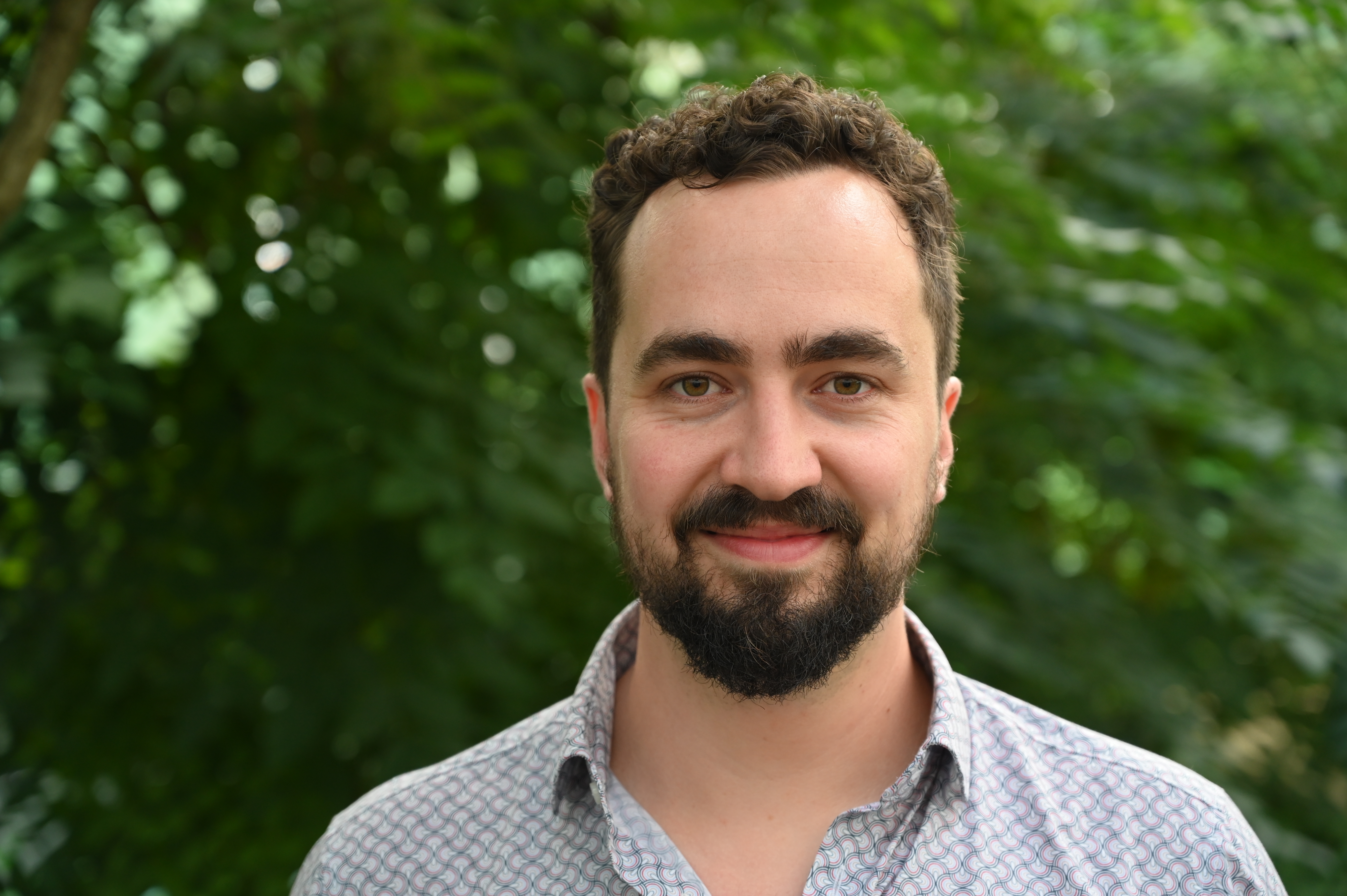
Karl Briegel, 29
Position
Co-founder of the start-up QTAS
Degree
Chemistry
At the start-up QTAS, Karl and his team are developing a new blood analysis device that will enable faster detection of the effectiveness of cancer treatments. The technology builds on optical NMR microscopy, whose functioning relies on quantum sensors in diamond.

Atomic defects in diamonds as quantum sensors
His research group has now developed a microscope that does something very similar to an MRI device in a hospital, the chemist continues, only on a very small scale and without producing a 3D image, but rather a 2D image of a sample placed on a specially prepared, tiny diamond chip. Unlike classical NMR methods, where the sample to be examined – in the case of an MRI, usually the entire body – is surrounded by a coil that detects the NMR signal by induction, the NMR microscope uses special quantum sensors for this purpose, Karl explains. “The diamond chip on which we position the sample that emits the signal contains an ensemble of NV centers.”
NV stands for "nitrogen vacancy" and refers to a defect in the diamond lattice. In diamond, which in its pure form consists of 100 percent carbon atoms, a nitrogen atom occupies a lattice site instead of a carbon atom. If, additionally, the neighboring lattice site is unoccupied, this is referred to as a nitrogen vacancy center, or NV center for short. The electronic structure of this atomic defect gives the NV center special properties that are crucial to the functioning of the NMR microscope. The spin of the NV center, which interacts with the oscillating magnetic field generated by the precessing nuclear spins in the sample, can be controlled using microwaves. Using special microwave pulse sequences – special “dance instructions” for the NV spin, as Karl calls them – one can make the NV center to listen as closely as possible to what the NMR signal is doing over several oscillations, the researcher explains. If, in addition, the NV center is excited with laser light, it fluoresces, with the intensity of the fluorescence depending on the state of the NV spin, he continues. “This ultimately allows us to translate the NMR signals into optical signals.” The original NMR signal can then be reconstructed from the fluorescence signal, which is filmed with a high-speed camera.
“We take one shot, 300 milliseconds, and have hundreds of thousands of NMR spectra”
Unlike an MRI device, which requires additional effort to determine where in the sample the signal detected by the coil originated, the NMR microscope automatically provides the localization required for imaging: “A single NV center in the diamond chip only measures the NMR signal in the immediate vicinity, which means we get a very localized signal coming from a very small bubble, that is, from the very small sample volume located directly above the NV center.” This also reflects the microscope's spatial resolution. This is in the range of a few micrometers, that is, on the order of magnitude of individual cells, Karl emphasizes, and because a camera is used, the many local NMR signals can all be measured simultaneously: “We take one shot, 300 milliseconds, and have hundreds of thousands of NMR spectra.” An MRI device would have to scan everything one after the other. “That's a huge bonus. It's extremely fast, with very high spatial resolution at the same time!”
Karl primarily uses the MRI comparison to highlight the NMR microscope's unique features. He makes clear, that the two technologies are not in direct competition with each other: “An MRI measures from a distance. You lie in this tube, there's a coil somewhere, and you still get an image of the middle of your head, for example.” Their microscope could not do that. “Our sensor measures very locally. For imaging, the sample is placed on the diamond chip.”
With QTAS, Karl and his colleagues now want to place blood samples from cancer patients onto the diamond chip and use the new technology to detect and count individual tumor cells in these blood samples. While his doctoral thesis focused on demonstrating the basic principle of the technology, the aim now is to turn it into a real application that can be used in clinics and really help people, the researcher proclaims: “We’re going full steam ahead to achieve this goal of building this device!”
Karl did not have his sights set on working in experimental quantum physics from the outset. By the time he finished high school, he knew he wanted to study chemistry. During his bachelor's program in Innsbruck, he realized he was particularly interested in physical and theoretical chemistry, so he decided to transfer to the TU Munich for his master's degree because it offered specialization in these two areas. For a long time, he thought he would ultimately end up in theoretical chemistry, Karl remembers. “Until one day Dominik came into a lecture.” The young professor had just arrived in Munich to establish a new group and a new laboratory at TUM. Dominik Bucher explained to the students that he used quantum sensors to perform NMR. He didn't lecture for long, just introduced himself briefly and sketched what he was doing on the blackboard, Karl recalls, and said that if anyone was looking for a master's, Ph.D., or internship position, they should just get in touch with him – “Which is what I did pretty much right after the lecture!” says Karl with a laugh. The short pitch immediately convinced the young chemist, and his good feeling was confirmed. He followed up the several-week internship with his master's thesis and eventually his doctoral thesis. “I just got completely hooked.”
What he enjoyed so much about working in the group and finally about his Ph.D. was, on the one hand, the fact that he was building something with a clear practical application and, on the other hand: “That the day-to-day work is extremely varied!” They had to build everything themselves. Of course, they bought components, but it wasn't a ready-made microscope, Karl emphasizes. “Basically, it's a jumble of cables and components, where you do everything from 3D printing to circuit board design. A large part of my job was also programming the whole thing.”
From research to starting a business
With QTAS, the range of tasks has become even broader. “Initially, I was concerned about moving away from technology,” says the young founder. “But essentially, my world has expanded.” He is still heavily involved with technology and has to tinker with things in the lab, but at the same time, there are now other things to think about: contact with doctors, speaking with investors outside the university, regulatory issues, cost efficiency, and much more.
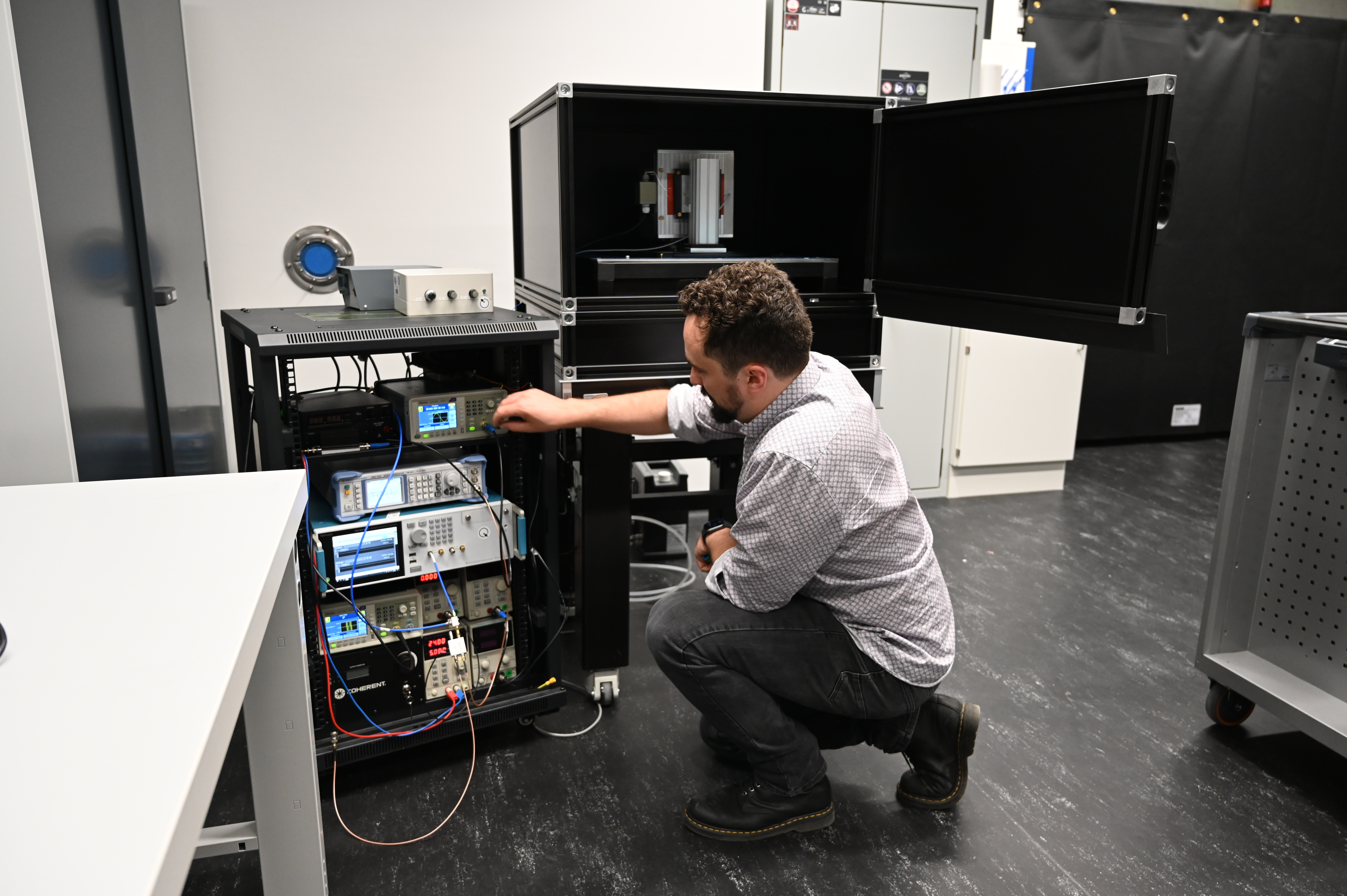
“We also spend a lot of time on very specific planning.” There is a long list of various to-dos and deadlines to keep in mind, so they have to prioritize and solve problems in a goal-oriented manner, Karl continues. That's why there are currently two workstations in the QTAS lab. While a large optical table in the middle of the room is used to optimize individual components and serves as a “workbench” where things can be tested and tried out, a relatively compact setup can be seen in another corner of the lab. “This is the device that will soon be going into the clinic,” says the founder, pointing to the significantly smaller optical table, where neither optics nor lasers are visible. They are hidden under a black box that is placed over the table and contains a silver-colored housing in which the diamond chip is installed. Not too many details can be seen from the outside. The electronics are stored next to the table in a waist-high cabinet. Unlike in research, this device no longer needs to be babysat by doctoral students, Karl jokes. “It's due to go into the clinic in three months, so everything has to be calibrated by then.”
Karl is grateful for how things turned out. A doctoral thesis on a topic that interests him greatly and a super nice team, and despite the heavy workload, he could always make time for friends, long walks with his fiancée and their two dogs, cooking evenings – “I'm a total foodie,” Karl admits – or trips to the mountains. Things are now moving forward in an exciting way with QTAS, says the young founder. “I'm very happy to have the opportunity to do this. I think it's going to be a really cool and wild ride!”
Published 26 September 2025; Interview 18 August 2025